X-ray artificial intelligence refers to machine learning systems trained to interpret X-ray crystallography data, automate structural analysis, and predict molecular configurations with unprecedented speed and accuracy. These AI models are transforming how researchers solve protein structures, accelerate drug discovery, and analyze complex biomolecular interactions that were once prohibitively time-consuming to decode manually.
The convergence of X-ray diffraction techniques and deep learning represents one of the most significant advances in structural biology since the invention of synchrotron radiation sources. Traditional X-ray crystallography requires expert crystallographers to spend weeks or months refining diffraction patterns, phasing data, and building atomic models. AI systems now complete portions of this workflow in hours, reducing bottlenecks in pharmaceutical pipelines and enabling high-throughput structural analysis that matches the pace of modern genomics and proteomics.
This technology matters because protein structure determines function. Understanding how enzymes fold, how antibodies bind to antigens, and how viral proteins interact with host cells depends on atomic-resolution imaging. The pharmaceutical industry invests billions annually in structure-based drug design, where knowing a target protein’s 3D architecture can mean the difference between a failed lead compound and a marketable therapeutic. AI-driven X-ray analysis has already contributed to solving previously intractable structures, including membrane proteins and large macromolecular complexes critical to disease mechanisms.
The integration of AI in molecular biology extends beyond crystallography into cryo-electron microscopy and small-angle X-ray scattering, creating a unified computational framework for structural determination. Researchers studying cell biology impact on therapeutic targets now routinely employ these tools to bridge molecular architecture with cellular function.
This article examines how X-ray AI systems function at a technical level, the distinct categories of algorithms deployed across structural biology workflows, and real-world applications reshaping drug development and molecular research in 2026.
What X-Ray AI Means for Modern Diagnostics and Research
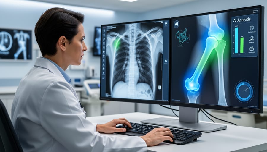
X-ray artificial intelligence comprises computational systems that apply machine learning algorithms to radiographic data, transforming how we interpret images from clinical chest radiographs to protein crystallography diffraction patterns. At its core, this technology leverages computer vision techniques and deep learning architectures to identify patterns that indicate disease states, structural anomalies, or molecular configurations, capabilities that extend far beyond simple image processing into genuine pattern recognition and predictive analysis.
These systems function by training neural networks on vast repositories of annotated X-ray images, learning to distinguish between normal tissue and pathology, between baseline crystal structures and novel conformations. Through iterative exposure to thousands or millions of examples, the algorithms develop an ability to recognize subtle features that might escape initial human observation: early-stage tumors in lung tissue, microfractures in bone scans, or slight variations in electron density maps that reveal protein binding sites crucial for drug development.
The technical foundation relies on several interconnected concepts that define how modern X-ray AI operates:
- Machine Learning
- A computational approach where algorithms improve performance on specific tasks through exposure to data rather than explicit programming, enabling systems to recognize complex patterns in radiographic images.
- Convolutional Neural Networks
- Specialized deep learning architectures designed to process grid-like data such as images, using layered filters to detect edges, textures, and higher-order features hierarchically.
- Radiomics
- The extraction of quantitative features from medical images, texture, shape, intensity distributions, that AI models analyze to predict clinical outcomes or biological characteristics.
- Computer-Aided Detection (CAD)
- Software systems that highlight potential areas of concern in radiographic images, flagging suspicious regions for radiologist or researcher review.
- Image Segmentation
- The process of partitioning an image into distinct regions or objects, allowing AI to isolate specific anatomical structures, lesions, or molecular features for targeted analysis.
What distinguishes contemporary X-ray AI from earlier computer-aided systems is the shift from rule-based algorithms to data-driven learning. Previous generations required programmers to manually define what constituted an abnormality; today’s deep learning models discover these patterns autonomously, often identifying novel biomarkers or structural correlations that advance scientific understanding.
In pharmaceutical development, this capability proves transformative. AI accelerates the analysis of X-ray crystallography data used to determine protein structures, compressing months of manual interpretation into hours of computational work. Biotechnology laboratories employ these systems to screen thousands of molecular configurations, identifying promising drug candidates with unprecedented speed. Clinical research facilities use X-ray AI to standardize imaging assessments across multi-site trials, reducing variability and improving the reliability of therapeutic efficacy measurements.
The augmentation model matters here: X-ray AI serves as a collaborative tool rather than a replacement for human expertise. Radiologists review AI-flagged findings within broader clinical context, while structural biologists validate AI-generated models against experimental constraints. This partnership combines computational processing power with human judgment, domain knowledge, and ethical oversight, a collaboration that has become standard practice in leading research institutions and pharmaceutical companies worldwide.
How X-Ray AI Systems Work
Image Acquisition and Preprocessing
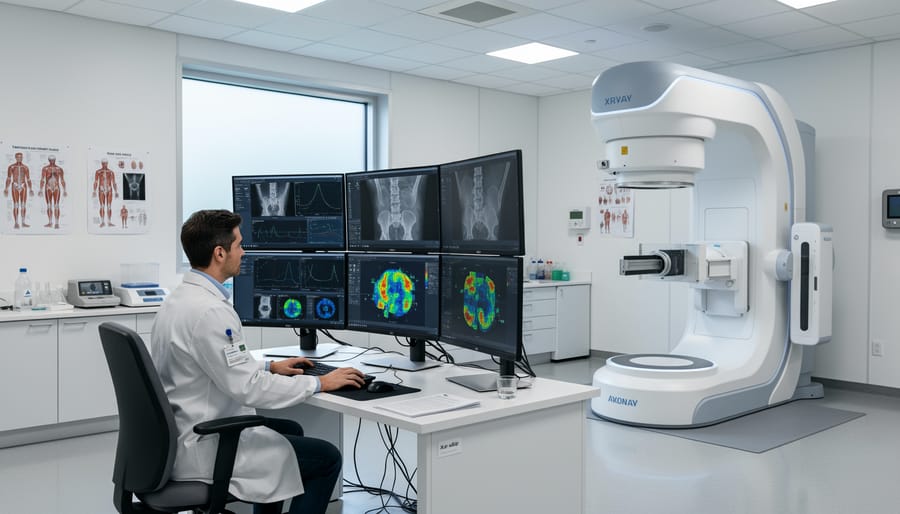
Digital X-ray images begin as raw sensor data captured by direct or indirect detector systems. In direct systems, X-ray photons convert immediately into electrical signals through amorphous selenium layers. Indirect systems use scintillator materials like cesium iodide to first convert photons into visible light, which photodiodes then transform into digital signals. Both methods produce grayscale images where pixel intensity values represent tissue density and atomic composition.
Before any AI analysis can occur, these raw images undergo systematic preprocessing to standardize input quality. Normalization scales pixel values to a consistent range, typically zero to one or negative one to positive one, eliminating variations caused by different imaging equipment, exposure settings, or detector sensitivities. This step is essential because neural networks trained on normalized data from multiple institutions cannot accurately process images with widely different intensity distributions.
Noise reduction algorithms filter out random signal fluctuations inherent in X-ray detection, particularly quantum noise from low photon counts and electronic noise from the detector circuitry. Techniques like Gaussian filtering, median filtering, or more advanced wavelet-based methods preserve structural details while suppressing artifacts that could mislead pattern recognition systems.
Additional quality enhancements may include contrast adjustment using histogram equalization, edge sharpening to emphasize anatomical boundaries, and artifact correction to remove beam hardening effects or scatter radiation. Some systems apply segmentation masks to isolate regions of interest, excluding irrelevant background areas that add no diagnostic value. These preprocessing steps transform variable raw radiographs into standardized, high-quality inputs optimized for consistent AI performance across diverse imaging conditions.
Neural Network Training and Pattern Recognition
Deep learning models for X-ray analysis rely on convolutional neural networks (CNNs) that process images through multiple layers of mathematical operations, progressively extracting features from simple edges to complex anatomical structures. Training begins with large datasets of annotated X-ray images where radiologists or domain experts have labeled specific features: fracture locations, tumor boundaries, protein crystal diffraction patterns, or molecular structures. The network processes these labeled examples repeatedly, adjusting internal parameters through backpropagation algorithms until it reliably predicts the correct labels. A typical diagnostic chest X-ray model might train on 100,000+ annotated images over several weeks of computational processing.
Supervised learning forms the foundation of most X-ray AI systems. Each training image serves as both input and answer key: the network sees the X-ray and simultaneously knows what it should find. Through iterative training cycles, the model learns which pixel patterns correlate with pathological findings or structural features. In molecular biology applications, networks trained on crystallography data develop specialized capabilities for recognizing diffraction patterns that indicate specific protein conformations, integrating seamlessly into data science pipelines that transform raw experimental data into actionable structural insights.
Validation ensures model reliability through holdout datasets the network never encountered during training. Researchers measure performance using metrics like sensitivity (ability to detect true positives) and specificity (avoiding false alarms), comparing AI performance against expert human interpretation. Models improve accuracy through transfer learning, where networks pre-trained on general radiographic images receive additional specialized training on specific conditions or molecular structures. This approach has proven particularly effective in structural biology, where automated electron density speeds protein structure determination by rapidly processing complex crystallographic data that would require days of manual analysis.
Analysis, Interpretation, and Output Generation
Once a neural network has learned to recognize patterns during training, it begins its real work: analyzing new X-ray images that practitioners submit for evaluation. The model receives a preprocessed image and passes it through multiple computational layers, each extracting increasingly complex features, from simple edges and textures to complete anatomical structures or crystallographic reflections.
As the analysis progresses, the system generates predictions about what it observes. For clinical applications, this might mean flagging potential pneumonia in lung regions or identifying hairline fractures in bone scans. In structural biology contexts, the AI interprets diffraction patterns to suggest electron density distributions or molecular conformations. Critically, the model doesn’t just provide binary yes-or-no answers. It assigns confidence scores to each prediction, typically expressed as percentages that indicate how certain the algorithm is about its findings.
These confidence metrics prove essential for practical deployment. A 98% confidence score on a detected nodule demands immediate attention, while a 62% score might prompt human review without triggering urgent protocols. The system typically highlights regions of interest directly on the image using bounding boxes, heat maps, or color overlays that guide the viewer’s attention to relevant areas.
Modern X-ray AI platforms integrate seamlessly with Picture Archiving and Communication Systems (PACS) and Laboratory Information Management Systems (LIMS), automatically receiving incoming images, processing them in seconds to minutes, and delivering structured reports. These reports appear within existing interfaces that radiologists and researchers already use, ensuring minimal workflow disruption while adding computational insight to human expertise.
Categories of X-Ray AI Applications
Clinical Diagnostic AI
Clinical diagnostic AI systems represent the most widely deployed category of X-ray artificial intelligence, trained to detect specific pathological features that indicate disease or injury. These algorithms analyze chest radiographs to identify pneumonia, tuberculosis, COVID-19, pulmonary nodules, and other respiratory conditions, often matching or exceeding radiologist accuracy in controlled studies. In orthopedic applications, AI detects fractures, bone density abnormalities, and joint degeneration with particular effectiveness in high-volume emergency department settings where rapid triage matters.
Oncology-focused systems scan mammograms and chest X-rays for early signs of malignancy, flagging subtle masses or architectural distortions that warrant further investigation. These tools operate as decision-support mechanisms rather than autonomous diagnostics, presenting findings alongside confidence scores that guide clinician interpretation. Modern implementations integrate seamlessly with PACS (Picture Archiving and Communication Systems), automatically analyzing incoming images and alerting radiologists to urgent findings.
The distinction between screening and diagnostic AI remains clinically significant: screening systems prioritize sensitivity to catch potential abnormalities, while diagnostic tools balance sensitivity with specificity to minimize false positives in confirmed case evaluation.
Structural and Molecular Research AI
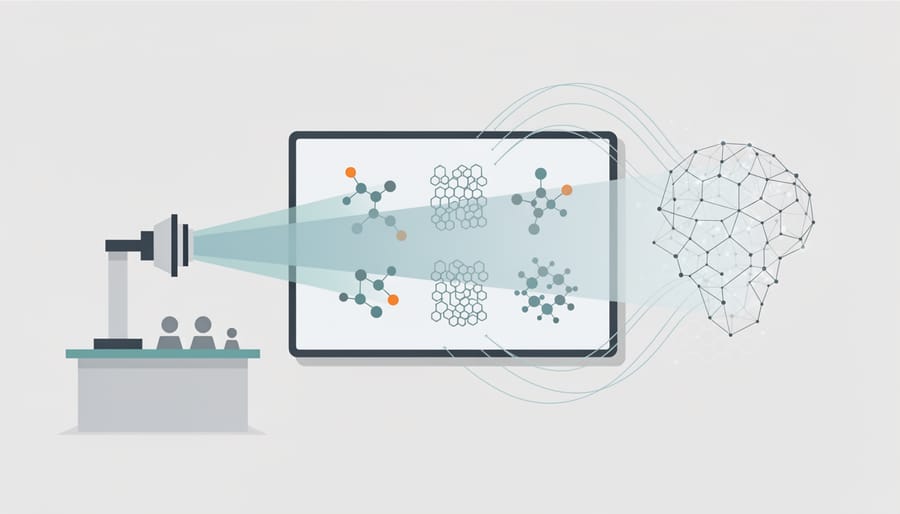
In X-ray crystallography and structural biology laboratories, artificial intelligence has fundamentally changed how researchers determine the three-dimensional structures of proteins and other biomolecules. Traditional crystallographic workflows require skilled crystallographers to interpret complex diffraction patterns and manually build molecular models into electron density maps, a process that can take weeks or months for challenging structures. AI systems now automate much of this analysis, recognizing patterns in diffraction data that indicate crystal quality, predicting phases from incomplete information, and building atomic models with accuracy approaching or sometimes exceeding manual interpretation.
Deep learning algorithms trained on thousands of solved protein structures can identify secondary structural elements, trace backbone chains through noisy density maps, and position amino acid side chains with remarkable precision. These tools prove especially valuable for large macromolecular complexes, membrane proteins, and low-resolution datasets where conventional methods struggle. In pharmaceutical research, this acceleration matters tremendously: rapidly determining the structure of a drug target protein or visualizing how candidate compounds bind to active sites shortens discovery timelines from months to days in some cases.
Beyond biomolecules, materials scientists deploy X-ray AI to characterize crystalline structures in new compounds, analyze defects in engineered materials, and identify phases in complex mixtures. The technology handles high-throughput screening at synchrotron facilities where thousands of samples generate data faster than human analysts could process, enabling researchers to explore chemical space more comprehensively and discover novel structures that advance both fundamental science and applied development programs.
Workflow Optimization and Triage Systems
In high-volume clinical environments and research laboratories processing thousands of X-ray images daily, AI-driven workflow optimization systems act as intelligent gatekeepers that route cases to the right specialist at the right time. These triage algorithms analyze incoming radiographs in real time, flagging critical findings like pneumothorax or suspected fractures for immediate review while deferring routine cases to standard queues. The result is reduced turnaround time for urgent diagnoses, often from hours to minutes, without requiring additional radiologist staffing.
Beyond prioritization, these systems automate quality assurance protocols that previously demanded manual oversight. AI validates image positioning, exposure levels, and anatomical coverage before images reach interpretation, rejecting substandard scans that would waste clinician time or require patient recalls. In research settings, particularly those conducting high-throughput crystallography experiments, similar AI tools pre-screen diffraction images to identify low-quality data early in the collection process. This automated quality control preserves valuable beam time and computational resources while maintaining rigorous data standards across large-scale structural biology projects.
How X-Ray AI Is Used Across Biomedical Research and Clinical Practice
Accelerating Drug Discovery Through Structural Analysis
AI-powered X-ray crystallography has fundamentally altered the pace of structure-based drug discovery. Traditional crystallography requires weeks of manual interpretation to convert diffraction patterns into accurate three-dimensional protein models. Deep learning systems now complete this analysis in hours, automatically resolving electron density maps and refining atomic coordinates with accuracy matching or exceeding expert crystallographers. This acceleration proves particularly valuable in pharmaceutical campaigns targeting complex proteins, kinases, GPCRs, and proteases, where understanding binding pocket geometry drives rational compound design.
The speed advantage compounds across discovery timelines. Research teams can now iterate through dozens of protein-ligand complexes during lead optimization, rapidly assessing how chemical modifications affect binding interactions. This structural feedback loop enables medicinal chemists to make data-driven decisions about molecular scaffolds, improving potency and selectivity before expensive synthesis campaigns. In oncology programs, including cancer immunotherapy development, AI-assisted structure determination has shortened the path from target identification to clinical candidate selection. Biopharmaceutical companies report reducing early discovery phases by 30-40%, with AI systems handling routine structural analysis while crystallographers focus on challenging cases requiring specialized expertise.
Enhancing Clinical Trials and Patient Stratification
X-ray AI transforms clinical trial design and execution by enabling precise patient selection and continuous disease monitoring through quantitative imaging analysis. During enrollment, AI systems analyze baseline radiographs to identify patients with specific disease characteristics, anatomical features, or biomarker patterns that match inclusion criteria, reducing screening failures and accelerating recruitment timelines critical to pharmaceutical development programs.
Throughout trial duration, AI-powered image analysis tracks subtle changes in tissue density, lesion size, or structural markers that indicate treatment response or disease progression. These quantitative measurements provide objective endpoints that supplement or replace subjective clinical assessments, strengthening statistical power and reducing required sample sizes. For oncology trials, AI detects millimeter-scale tumor changes weeks before they become clinically apparent, enabling adaptive trial designs that adjust dosing or patient cohorts based on early efficacy signals.
In precision medicine applications, X-ray AI stratifies heterogeneous patient populations into subgroups with distinct radiological phenotypes, revealing which molecular profiles respond best to specific interventions. This imaging-based stratification identifies biomarker-defined populations for targeted therapies, particularly in respiratory diseases, bone disorders, and metastatic cancers where radiographic features correlate with underlying molecular mechanisms and treatment outcomes.
Supporting Biopharmaceutical Quality Control
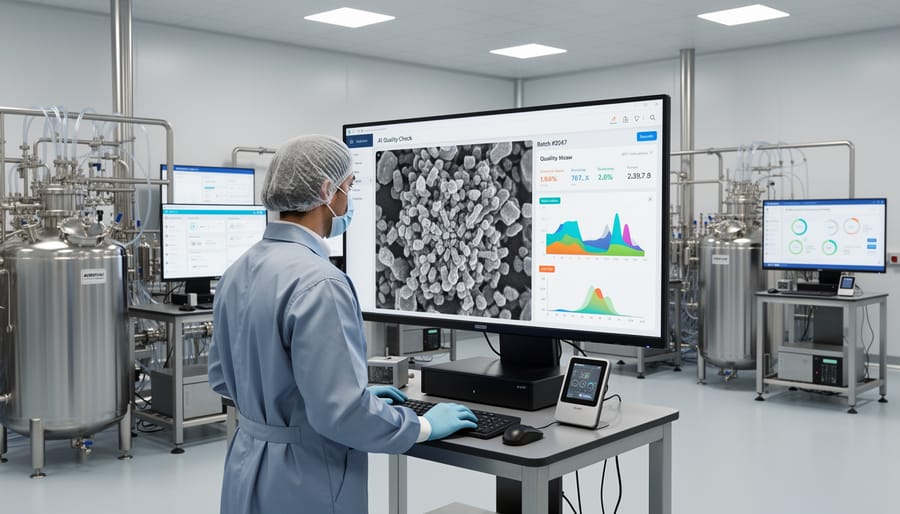
X-ray AI has become integral to pharmaceutical manufacturing quality control, where it addresses the stringent inspection requirements for sterile injectables, prefilled syringes, and vial-based biologics. Automated X-ray inspection systems equipped with machine learning algorithms scan every container on production lines at speeds exceeding 600 units per minute, detecting fill-level deviations, foreign particulate matter, and container defects that human inspectors might miss during visual checks.
In biologics manufacturing, X-ray AI identifies glass fragments, metal particles, and rubber stopper debris with detection thresholds below 50 micrometers, critical for patient safety in high-value therapeutic proteins and monoclonal antibodies. These systems analyze radiographic density variations to distinguish acceptable air bubbles from unacceptable contaminants, reducing false rejection rates that waste expensive product while maintaining zero-defect release standards.
Beyond container inspection, X-ray AI monitors lyophilized cake structure in freeze-dried products, verifying proper reconstitution characteristics and detecting collapsed or incomplete drying that would compromise stability. The technology also validates proper component assembly in combination products like autoinjectors, confirming needle placement and spring mechanism integrity. This automated quality assurance provides real-time production feedback, generates compliance documentation for regulatory submissions, and enables manufacturers to meet FDA and EMA requirements for pharmaceutical quality systems while maintaining the throughput necessary for commercial-scale biopharmaceutical production.
Accuracy, Validation, and Collaborative Implementation
Deploying X-ray AI systems in clinical and research environments demands rigorous validation protocols that extend far beyond initial development benchmarks. Industry leaders recognize that a model demonstrating 95% accuracy in controlled testing conditions must prove itself against diverse patient populations, varied imaging equipment, and real-world data quality inconsistencies before earning regulatory approval or research implementation. The FDA’s regulatory pathway for AI-based diagnostic devices now requires continuous performance monitoring post-deployment, acknowledging that algorithms trained on historical datasets can drift in accuracy as patient demographics and imaging technologies evolve.
Validation frameworks in biopharmaceutical research laboratories incorporate multiple verification layers. When pharmaceutical companies deploy AI for protein crystallography analysis, they compare algorithmic outputs against gold-standard manual interpretations from experienced crystallographers across hundreds of test structures. This parallel validation reveals not just overall accuracy rates but specific failure modes, certain crystal symmetries, resolution limits, or sample preparation artifacts where human oversight remains essential. Teams document these boundaries explicitly in standard operating procedures, establishing clear handoff points between automated screening and expert review.
The collaborative implementation model treats AI as an augmentation tool rather than autonomous decision-maker. In clinical radiology departments, AI flags suspicious findings and assigns urgency scores, but radiologists retain final diagnostic authority and frequently catch nuances the algorithm misses, subtle asymmetries, clinical history correlations, or rare presentations outside the training distribution. This human-in-the-loop approach captures discrepancies in systematic error logs that feed back into model retraining cycles. Leading research institutions apply similar frameworks to structural biology workflows, where AI accelerates initial structure determination but crystallographers validate bond angles, electron density interpretations, and conformational states that determine biological function.
Continuous improvement cycles depend on structured expert feedback mechanisms. Pharmaceutical AI vendors now build annotation interfaces directly into deployment platforms, allowing researchers to flag incorrect predictions, confirm accurate calls, and contribute challenging cases to expanding training datasets. This creates a virtuous cycle where domain expertise continuously refines algorithmic performance, particularly important as molecular medicine trends introduce novel disease presentations and therapeutic targets not represented in historical training data. The most successful implementations measure success not by automation rates but by the quality of human-AI collaboration, faster time to insight, reduced expert fatigue on routine cases, and enhanced focus on complex problems requiring scientific judgment.
Common Questions About X-Ray AI
Many professionals considering X-ray AI implementation face similar questions about performance, feasibility, and practical deployment. These frequently asked questions address the most common concerns raised by clinical laboratories, research institutions, and biopharmaceutical companies evaluating this technology.
How accurate are X-ray AI systems compared to human radiologists?
Top-performing X-ray AI systems match or exceed human radiologists for specific tasks like detecting lung nodules or fractures, often achieving accuracy rates above 90% when trained on large, high-quality datasets. However, accuracy varies significantly by condition type, image quality, and population demographics, which is why deployment combines AI with human oversight rather than replacing expert interpretation.
Can AI detect conditions that human experts miss?
Yes, AI can identify subtle patterns in radiographic images that escape human attention, particularly early-stage pathologies or rare conditions underrepresented in training. Studies demonstrate that AI excels at detecting micro-fractures, small pulmonary nodules, and diffraction pattern anomalies in crystallography that humans might overlook during high-volume screening.
What data is required to train an X-ray AI system?
Effective training requires thousands to millions of annotated X-ray images representing diverse patient populations, imaging equipment, and pathological variations. For specialized applications like protein crystallography analysis, datasets need expert-labeled diffraction patterns covering multiple crystal systems and experimental conditions to ensure robust performance across research scenarios.
How are false positives managed in clinical settings?
Clinical deployments incorporate confidence scoring that flags uncertain cases for human review, adjustable sensitivity thresholds tailored to specific use cases, and secondary validation workflows where radiologists confirm AI findings before clinical decisions. This layered approach maintains high sensitivity while controlling false positive rates through expert oversight.
Beyond accuracy metrics, implementation considerations weigh heavily on institutional decisions. Regulatory approval status varies by jurisdiction and intended use. In the United States, multiple X-ray AI systems have received FDA clearance for specific diagnostic tasks, while European CE marking covers others. Research applications in crystallography and materials analysis typically fall outside medical device regulations but still require validation against established benchmarks.
Cost structures span initial licensing fees, ongoing computational infrastructure, integration with existing PACS and laboratory information management systems, and staff training. Small clinical laboratories might spend tens of thousands annually for cloud-based solutions, while large pharmaceutical research facilities investing in on-premise systems face six-figure implementation costs. However, these expenses must be weighed against efficiency gains, faster turnaround times, and improved diagnostic or analytical throughput that directly impact research timelines and patient outcomes.
Data privacy and security protocols require careful attention, particularly when training models on patient images or proprietary research data. Institutions must ensure compliance with HIPAA, GDPR, or other relevant regulations, establish clear data governance policies, and verify that vendor agreements protect confidential information. For biopharmaceutical companies, this extends to safeguarding competitive intelligence embedded in structural biology datasets used for drug discovery programs.
X-ray artificial intelligence stands at the intersection of computational innovation and scientific discovery, fundamentally reshaping how researchers and clinicians extract meaning from radiographic data. This technology exemplifies the broader transformation occurring across scientific disciplines as machine learning systems augment human analytical capabilities with unprecedented speed and precision.
The most successful implementations of X-ray AI follow a collaborative model rather than a replacement paradigm. Deep learning algorithms excel at processing vast volumes of imaging data and identifying subtle patterns beyond human perceptual limits, while experienced professionals provide critical context, validate findings, and make nuanced decisions that require clinical judgment or experimental insight. This partnership approach maximizes accuracy, maintains scientific rigor, and preserves the irreplaceable value of domain expertise.
For molecular biology researchers, X-ray AI accelerates protein crystallography analysis and structural determination workflows that directly support drug discovery pipelines. In clinical laboratories and biopharmaceutical facilities, these systems enhance diagnostic accuracy, streamline quality control processes, and enable precision medicine approaches that depend on reliable imaging biomarkers. The technology’s continued evolution promises tighter integration with laboratory information systems, expanded capabilities in complex structural analysis, and increasingly sophisticated decision support across research and clinical applications.
As institutions evaluate AI adoption, the focus should remain on systems that enhance rather than automate professional expertise, ensuring that computational power serves scientific advancement while maintaining the collaborative human oversight essential to rigorous research and patient care.